Top > About Us > News Letter > Department of Radiology Achieves World's Lowest Exposure Dose for Infants
Department of Radiology Achieves World's Lowest Exposure Dose for Infants
From The University of Tokyo Hospital to the World
Department of Radiology
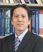
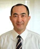
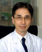
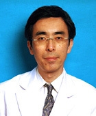
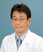
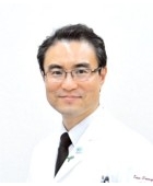
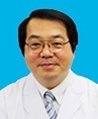
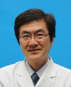
Osamu Abe, Professor and Chairman
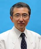
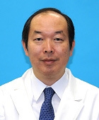
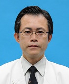
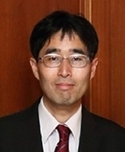
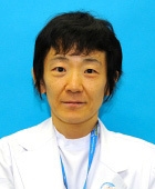
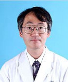
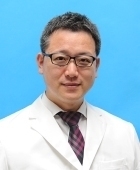
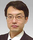
Written by Eriko Maeda, Project Assistant Professor
The Department of Radiology handles the full spectrum of radiological examination and treatment at the University of Tokyo Hospital. The department is comprised of the division of diagnostic radiology, which undertakes accurate systemic diagnostic imaging (CT, MRI, etc.), systemic angiography, and endovascular treatment; the division of nuclear medicine, which undertakes PET and RI examination; and the division of radiotherapy, which undertakes radiotherapy for cancer and vascular malformation. This issue introduces the challenges taken on by the division of diagnostic radiology to achieve the world’s lowest level of radiation exposure in cardiac CT examination.
1. Clinical diagnostic radiology
In modern medicine, the diagnosis and follow-up of many diseases depend on diagnostic imaging. While radiologists have few opportunities to see patients face-to-face, they are deeply engaged in team care as the only specialists who diagnose all diseases affecting the entire body through imaging.
When requests for examination are received from clinical departments, we first examine appropriate test methods to match the conditions, including the necessity of contrast medium in CT; the injection rate and scanning timing if a contrast medium is used; and what types of images should be taken in MRI (diagnosis is performed by comparing images that are taken using various methods, such as T1-weighted scans and diffusion-weighted images). During tests, radiologists are focused on safe examination with low exposure. Following scanning, they proceed to the interpretation process, in which radiologists check the images for abnormalities through systematic examination of all organs. If an abnormality is found, detailed findings are presented, and around 10 disease candidates are listed. Subsequently, the list is narrowed down to the three or so that seem the most likely. After prioritizing items in the shortlist, the interpretation of systemic findings and of abnormal findings are described in writing, and communicated to the relevant clinical departments as the final step in the process. Radiologists participate in almost all clinical conferences, including those of the cancer board, and provide inputs as imaging specialists.
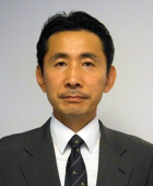
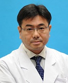
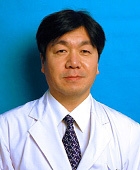
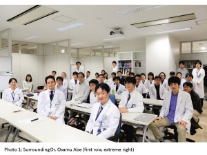
The University of Tokyo Hospital has more than 20 board-certified diagnostic radiologists. The division of diagnostic radiology has approximately 30 radiologists, including graduate students and medical interns. In October 2016, we welcomed new professor Dr. Osamu Abe, and we are renewing our efforts in clinical practice, education, and research (Photo 1).
2. Reduced exposure in cardiac CT
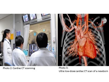
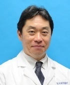
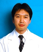
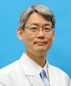
There are extremely large differences among facilities in terms of the image quality of cardiac CT and the exposure doses involved, depending on the form of participation by radiologists. In our department, experienced diagnostic radiologists specializing in cardiovascular imaging, led by Project Assistant Professor Maeda, have performed cardiac CT on patients ranging from newborn babies to the elderly, while at the same time conducting ongoing research toward reduced exposure (Photo 2).
Patients with congenital heart diseases must undergo a series of examinations accompanying radiation exposure, including catheterized examination and treatment in addition to CT scans, and infants, in particular, are highly sensitive to radiation. This is why efforts have been made to minimize the exposure dose in CT. We have succeeded in taking high-quality cardiac CT images of infants with the lowest-level dose in the world (approx. 0.2 mSv) by applying full iterative reconstruction to the state-of-the-art 320-slice CT. This is on the level of one-tenth to one-hundredth of the dose received in infant cardiac CT at a normal hospital, and is equivalent to one routine chest x-ray (or because the exposure dose in routine chest radiography is also low at our hospital, equivalent to 10 scans), or to a round-trip flight between Japan and the U.S.
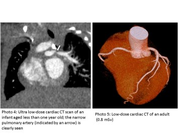
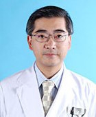
While 320-slice CT scans have become popular in Japan and overseas, images at our hospital are remarkably clear despite the significantly low dose. The main reason for this is that because they have had considerable experience in the diagnosis of patients with complicated congenital heart disease, our radiologists have developed an adequate understanding of images required in clinical settings through communication with pediatricians and thoracic surgeons, and have thereby developed methods for using contrast mediums in accordance with various hemodynamic patterns. Because the heart of an infant measures just several centimeters, images are required to have an accuracy of between half to one millimeter. Furthermore, the rate of their heartbeat is fast, while blood flows vary depending on the congenital heart disease, making imaging difficult in and of itself, even before examining any possible reduction in the exposure dose. The extreme level of specialization is close to that of macro photographers or astrophotographers. Because our hospital has established basic infrastructure to cope with the highest level of difficulty, we are able to acquire images with sufficient clarity at reduced doses. Moreover, each single test can be finished without the fear of retaking, as failure is extremely rare. Because our radiologists have become accustomed to working with many images, they can perform a proper diagnosis, which contributes to an even lower dose, thereby forming a virtuous circle. This is how our hospital has achieved routine examination at a low dose that no other facility in the world can match (Photos 3 and 4). Another element that has contributed to our success is that we are able to use state-of-the-art models and image processing programs as part of joint research conducted with manufacturers.
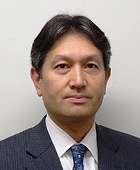
Lower dose does not always mean better imaging. Just as is the case with underexposure in a camera, too low a dose compromises image quality. Without the requisite experience, it is difficult to reduce the dosage when working with actual patients. We therefore apply findings collected through infant cardiac CT to adults, thereby achieving a reasonable reduction in exposure (Photo 5). We invite you to come and receive radiological examination at our hospital with peace of mind.
Departments/Divisions
Pediatrics, Children's Medical Center
Titles
M.D. , Ph.D.
Expertise/Specialties
Pediatrics, Child Neurology, Perinatal and Neonatal Medicine
Research Interests
1) Perinatal brain injury 2) Environmental effects of development
3) Congenital cytomegalovirus infection 4) Developmental disorders
5) Subacute Sclerosing Panencephalitis 6) Public health screening system
Languages
Japanese/ English
See More
Departments/Divisions
Urology and Andrology
Titles
M.D. , Ph.D.
Expertise/Specialties
Robotic Surgery, Laparoscopic Surgery, Endoscopic Surgery, Kidney Cancer, Urinary Tract Cancer, Bladder Cancer, Prostate Cancer, Testicular Cancer, Adrenal Tumor, Renal failure, Peritoneal Dialysis
Research Interests
Molecular Biology, Genome Research
Languages
See More
Departments/Divisions
Clinical Research Governance
Titles
Ph.D.
Expertise/Specialties
Clinical Reseach, GCP, regulation
Research Interests
Quality Management
Languages
Japanese / English
See More
Departments/Divisions
Department of Neurosurgery
Titles
M.D. , Ph.D.
Expertise/Specialties
Acoustic neuroma surgery, Arteriovenous malformation surgery, Skull base meningioma surgery, vertebrobasilar aneurysm surgery
Research Interests
Functional preservation of brain, brainstem and cranial nerves, surgical simulation with 3DCG, functional restoration by brain-machine interface (BMI), neural regeneration by recruitment of endogenous progenitors
Languages
Japanese/ English
See More
Departments/Divisions
Pharmaceutical Department
Titles
Ph.D.
Expertise/Specialties
pharmacy
Research Interests
clinical and molecular pharmacology / toxicology, and systems-biology / pharmacology / toxicology
Languages
Japanese / English
See More
Departments/Divisions
Department of Blood Transfusion
Titles
M.D., Ph.D.
Expertise/Specialties
Transfusion Medicine, Respiratory Medicine, Allergy and Clinical Immunology
Research Interests
Mechanism of transfusion complications, Haemovigilance
Languages
Japanese / English
See More
Departments/Divisions
Department of Pediatric Surgery
Titles
M.D. , Ph.D.
Expertise/Specialties
Pediatric Surgery, Pediatric Minimally Invasive Surgery, Pediatric Oncology, Pediatric Urology, Pediatric HBP Surgery
Research Interests
Pediatric Hepatobiliary disease, Pediatric MIS, Transplantation Immunology
Languages
Japanese/English
See More
Departments/Divisions
Central Supply Service
Titles
M.D. , Ph.D.
Expertise/Specialties
General Surgery, Operative Medicine, Nutrition Support
Research Interests
Surgical Nutrition and Metabolism, Gut Immunity
Languages
Japanese, English
See More
Departments/Divisions
Department of Gastroenterology; Department of Endoscopy and Endoscopic Surgery
Titles
M.D. , Ph.D.
Expertise/Specialties
Gastroenterology, Hepatology (Hepatitis, Hepatocellular Carcinoma), Pancreatology
Research Interests
Viral hepatitis, Hepatocarcinogenesis, NASH, Oxidative stress
Languages
Japanese / English
See More
Departments/Divisions
Allergy and Rheumatology
Titles
M.D. , Ph.D.
Expertise/Specialties
Rheumatology, Internal Medicine
Research Interests
Clinical Immunology, Basic Immunology
Languages
Japanese, English
See More
Departments/Divisions
Department of Psychosomatic Medicine
Titles
M.D. , Ph.D.
Expertise/Specialties
Psychosomatic Medicine
Research Interests
Research on development of treatment for eating disorders and on development of mobile tools for assessment and treatment for life-style related disorders and mood disorders using EMA and EMI methods.
Languages
Japanese / English
See More
Departments/Divisions
Neuropsychiatry, Medical Community Network and Discharge Planning
Titles
M.D. , Ph.D.
Expertise/Specialties
Clinical Psychiatry, Early intervention and rehabilitation for schizophrenia, Community mental health
Research Interests
Neuroimaging in psychiatric disorders, Mental health and neuroscience in adolescence
Languages
Japanese / English
See More
Departments/Divisions
Infection Control and Prevention Service/Department of Infection Control and Prevention Graduate School of Medicine
Titles
M.D. , Ph.D.
Expertise/Specialties
Infection Control and Prevention Service/ Hepatocellular carcinoma, Liver diseases, Viral hepatitis, Liver diseases
Research Interests
Mechanism of hepatocarcinogenesis and relarion between carcinogenesis and mitochondria damage
Languages
Japanese/English
See More
Departments/Divisions
Ophthalmology
Titles
M.D., Ph.D
Expertise/Specialties
Glaucoma, Neurobiochemistry, Ocular Pharmacology
Research Interests
・the analysis of the risk factor activating glaucoma
・the study of the surgical outcomes of glaucoma
・the elucidation to the pathology of increasing intraocular pressure through bioactive lipids
・the development of new drops for glaucoma
Languages
Japanese, English
See More
Departments/Divisions
Breast and Endocrine Surgery
Titles
M.D. , Ph.D.
Expertise/Specialties
surgery on primary breast cancer, systemic treatment on metastatic breast cancer
Research Interests
epigenetic change which is critical for breast cancer development, transcriptional regulation of estrogen receptor alpha (ERα)in breast cancer
Languages
Japanese/English
See More
Departments/Divisions
Division of Nephrology and Endocrinology, Department of Hemodialysis and Apheresis
Titles
M.D. , Ph.D.
Expertise/Specialties
Chronic kidney disease, Acute kidney injury, End stage kidney disesae, Hemodialysis, Nephrotic syndrome, Renal anemia, Atypical hemolytic uremic syndrome
Research Interests
Oxygen metabolism of the kidney, immunological kidney injury, epigenetics, renal anemia
Languages
Japanese/English
See More
Departments/Divisions
Department of Pain and Palliative Medicine
Titles
M.D. , Ph.D.
Expertise/Specialties
Pain Medicine, Palliative Medicine, Anesthesiology, Critical Care Medicine, Medical Engineering
Research Interests
Pain Medicine, Palliative Medicine, Cognitive Neuroscience, Health Literacy
Languages
Japanese
See More
Departments/Divisions
Geriatric Medicine
Titles
M.D. , Ph.D.
Expertise/Specialties
geriatric medicine, gerontology
Research Interests
Pharmacotherapy and its safety in the elderly. Gender difference in geriatric medicine.
Languages
Japanese, English
See More
Departments/Divisions
Department of Hematology and Oncology
Titles
M.D. , Ph.D.
Expertise/Specialties
Hematological malignancies
Research Interests
Leukemia
Languages
Japanese, English
See More
Departments/Divisions
Department of Cardiovascular Surgery & Department of Cooperative Unit of Medicine and Engineering Research
Titles
M.D. , Ph.D.
Expertise/Specialties
Adult Cardiac Surgery, Minimally Invasive Cardiac Surgery, Robotic Cardiac Surgery, Mitral Valve Plasty, Off-pump Coronary Artgery Bypass Surgery, Ventricular Assist Device, Heart Transplantation
Research Interests
Surgical Treatment of End-stage Heart Failure (Ventricular assist device, Heart transplantation, Mitral complex plasty), Device Development for Minimally Invasive Surgery, Regenerative Medicine
Languages
Japanese/English
See More
Departments/Divisions
Plastic Reconstructive and Aesthetic Surgery
Titles
M.D., Ph.D.
Expertise/Specialties
Reanimation of Established Facial paralysis, Reconstruction of Facial Deforimites, Wound Healing
Research Interests
Microsurgery, Facial Paralysis, Ageing
Languages
Japanese/English
See More
Departments/Divisions
Department of Acute Medicine
Critical Care and Emergency Medical Center/Emergency Room, Intensive Care Unit
Titles
M.D., Ph.D
Expertise/Specialties
emergency medicine, critical care medicine, intensive care medicine, disaster medicine, mass gathering medicine
Research Interests
acuity, triage, monitoring, emergency medical service system, disaster medical response system
Languages
English /Japanese
See More
Departments/Divisions
Department of Rehabilitation Medicine, Rehabilitation Center
Titles
M.D. , Ph.D.
Expertise/Specialties
Pediatric Rehabilitation, Rehabilitation and Prosthetics/Orthotics for Congenital Limb Malformation, Rehabilitation for Bone Dysplasias
Research Interests
Pediatric Rehabilitation, Rehabilitation for Disabled Children, Motion Analysis
Languages
Japanese / English
See More
Departments/Divisions
Center for Epidemiology and Preventive Medicine
Titles
M.D. , Ph.D.
Expertise/Specialties
Gastroenterology, Preventive Medicine
Research Interests
Oncology, Epigenetics, Molecular biology, Differentiation and Cancer
Languages
Japanese, English
See More
Departments/Divisions
Neurosurgery
Titles
M.D. , Ph.D.
Expertise/Specialties
Neurosurgery
Research Interests
Surgery of cerebrovascular diseases, Surgery of benign brain tumors, Experimental cerebral ischemia
Languages
Japanese/English
See More
Departments/Divisions
Department of Radiology
Division of Diagnostic Radiology
Titles
M.D., Ph.D
Expertise/Specialties
General diagnostic radiology, neuroradiology, interventional radiology
Research Interests
Voxel-based analysis, voxel-based morphometry, diffusion magnetic resonance imaging,
functional magnetic resonance imaging
Languages
Japanese/English
See More
Departments/Divisions
Stomach and Esophageal Surgery, Cancer Resource Center
Titles
M.D. , Ph.D.
Expertise/Specialties
Abdominal Surgery, General Surgery, Cancer Patients' Care
Research Interests
Gastric Carcinogenesis, Stem Cell and Carcinogenesis, Cancer Biomarker, Cancer Immunology, Growth Factor, Development
Languages
English, Japanese
See More
Departments/Divisions
Orthopaedic Surgery and Spinal Surgery
Titles
M.D. , Ph.D.
Expertise/Specialties
Joint surgery, rheumatoid arthritis, osteoporosis
Research Interests
Bone and cartilage biology, arthritis
Languages
Japanese, English
See More
Departments/Divisions
Dermatology
Titles
M.D. , Ph.D.
Expertise/Specialties
Scleroderma
Research Interests
Scleroderma, B lymphocytes, Autoimmunity
Languages
Japanese/English
See More
Departments/Divisions
Colorectal Surgery; Vascular Surgery
Titles
M.D., Ph.D
Expertise/Specialties
General Surgery, Gastrointestinal Surgery, Colorectal Surgery, Laparoscopic Surgery, Robotic Surgery, Minimally Invasive Surgery, Chemotherapy, Gastrointestinal Endoscopy, Colorectal Disease, Anorectal Disease, Colorectal Cancer, Inflammatory Bowel Disease, Diverticular Disease, Colorectal Polyp, Vascular Surgery, Abdominal Aortic Aneurysm, Thoracic Aortic Aneurysm, Endovascular Aneurysm Repair, Thoracic Endovascular Aortic Repair, Distal Bypass, Critical Limb Ischemia, Takayasu's Disease, Buerger Disease, Pancreatoduodeal Artery Aneurysm, Popliteal Entrapment Syndrome, Behçet's Disease, Carotid Endarterectomy, Hemodialysis, Peripheral Artery Aneurysm, Segmental Arterial Mediolysis, Deep Vein Thrombosis
Research Interests
Surgical Oncology, Vascular Surgery
Languages
Japanese/English
See More
Departments/Divisions
International Medical Center
University of Tokyo Tissue Bank
Artificial Organ and Transplantation Division, Department of Surgery
Titles
M.D., Ph.D., F.A.C.S.
Expertise/Specialties
Surgery, Heaptology, Liver Transplantation, Tissue Transplantation, Medical Education
Research Interests
Liver Disease, Liver Trasnplantation, Organ Transplantation, Donor Safety in Living Liver Donor, Tissue Transplantation, Tissue Banking, Cyropreservation of homograft, Medical Education, Surgical Training, Medical Care for Foreign Patients in Japan, Multicultural Resource for Health Care, Cross border clinical medicine
Languages
Japanese / English
See More
Departments/Divisions
Respiratory Medicine
Titles
M.D. , Ph.D.
Expertise/Specialties
Respiratory Medicine
Research Interests
The mechanism of respiratory diseases including COPD, asthma and pulmonary fibrosis
Languages
Japanese/English
See More
Departments/Divisions
University Hospital Medical Information Network Center Department of Heath Communication, School of Public Health, Faculty of Medicine, the University of Tokyo
Titles
M.D., Ph.D.
Expertise/Specialties
health communication, health informatics
Research Interests
Interpersonal and media-based health communication
Languages
Japanese/English
See More
Departments/Divisions
Department of Diabetes and Metabolic Diseases
Titles
M.D., Ph.D.
Expertise/Specialties
Diabetes, Metabolism, Obesity, Nutrition,
Research Interests
Pathogenesis of type2 diabetes, insulin resistance, adiponectin
Languages
Japanese / English
See More
Departments/Divisions
Clinical Research Support Center
Titles
M.D. , Ph.D.
Expertise/Specialties
Clinical Pharmacology, Neurology
Research Interests
Clinical Pharmacology, Neurology
Languages
Japanese, English
See More
Departments/Divisions
Otorhinolaryngology and Auditory and Voice Surgery
Titles
M.D. , Ph.D.
Expertise/Specialties
Otology, Audiology, Neurotology
Research Interests
Cochlear implant, hearing loss, regeneration, anti-aging
Languages
Japanese, English
See More
Departments/Divisions
Pathology
Titles
M.D. , Ph.D.
Expertise/Specialties
Gastrointestinal pathology
Research Interests
The pathology and molecular biology of gastrointestinal tumor
Languages
Japanese, English
See More
Departments/Divisions
Environment, Health and Safety Office
Titles
M.D., Ph.D.
Expertise/Specialties
Neurology
Research Interests
Clinical Neurology, Quality and Safety in Hospital Practice
Languages
Japanese, English
See More
Departments/Divisions
Obstetrics and Gynecology, Perinatal Center
Titles
M.D. , Ph.D.
Expertise/Specialties
Perinatal care for both normal and abnormal antepartum, labor, delivery, fetus and newborn, puerperium
Research Interests
Reproductive immunology / Perinatology / Reproductive Endocrinology
Languages
Japanese / English
See More
Departments/Divisions
Department of Diabetes and Metabolic Diseases
Titles
M.D. , Ph.D.
Expertise/Specialties
Internal Medicine, Diabetes, Metabolism, Nutrition, Obesity, Metabolic Syndrome, Diabetic Complications, Atherosclerosis, Insulin Resistance, Adipokines, Nuclear Receptors, Epigenetics, GWAS, Sportology, Anti-Aging Medicine
Research Interests
The mechanisms by which obesity results in insulin resistance, atherosclerosis and short life
Languages
Japanese / English
See More
Departments/Divisions
Stomach and Esophageal Surgery, Breast and Endocrine Surgery
Titles
M.D. , Ph.D.
Expertise/Specialties
Upper GI surgery, Esophageal Cancer, Gastric Cancer
Research Interests
surgical procedure and oncology of esophageal and gastric cancer
Languages
Japanese and English
See More
Departments/Divisions
Anesthesiology and Pain Relief Center
Titles
M.D. , Ph.D.
Expertise/Specialties
Anesthesiology, Critical care medicine, Respiratory care, Operative medicine, Pain medicine
Research Interests
Acute lung injury, Mechanical ventilation, acute inflammatory response, Mechanism of general anesthesia, Modulation of pain
Languages
Japanese/ English
See More
Departments/Divisions
Department of Child Psychiatry
Titles
M.D. , Ph.D.
Expertise/Specialties
Child and Adolescent Psychiatry, Tourette Syndrome and Other Tic Disorders, Obsessive-Compulsive Disorder (OCD), Attention-Deficit/Hyperactivity Disorder (ADHD), Autism Spectrum Disorder (ASD), School Mental Health
Research Interests
Phenomenology, Pathogenesis and Intervention of Tourette Syndrome and Comorbid Disorders Including OCD and ADHD
Languages
Japanese/English
See More
Departments/Divisions
Gynecologic Surgery
Titles
Professor
Expertise/Specialties
Reproductive medicine, laparoscopic surgery, assisted reproductive technology
Research Interests
Pathogenesis and management of endometriosis, Ovarian physiology/ pathology
Languages
Japanese / English
See More
Departments/Divisions
Department of Clinical Laboratory
Titles
M.D. , Ph.D.
Expertise/Specialties
Laboratory Medicine, Clinical Hematology, Thrombosis and Hemostasis
Research Interests
Platelet Biology, Vascular Biology, Bioactive Lipids
Languages
Japanese/ English
See More