Top > About Us > News Letter > Fused technique of gastroenterology and gastrointestinal surgery – NEWS from UTokyo
Fused technique of gastroenterology and gastrointestinal surgery – NEWS from UTokyo
Department of Endoscopy and Endoscopic Surgery, Manager and Associate Professor, Mitsuhiro Fujishiro, M.D. & Ph.D.
(1) Introduction to the Department of Endoscopy and Endoscopic Surgery
The Department of Endoscopy and Endoscopic Surgery, which started as a part of the Department of Clinical Laboratory, has been independent as one of the Central Clinical Facilities since April 1997 because of the increasing expertise and importance of endoscopic medical care. In our department, nearly 20,000 endoscopic examinations and treatments are performed every year, including gastrointestinal endoscopy, such as “gastric cameras” (Table 1). Although our full-time employees are only an associate professor and an assistant professor, who are both gastroenterologists, other physicians in the Departments of Gastroenterology, Stomach and Esophageal Surgery, Colon and Rectal Surgery, Hepatobiliary and Pancreatic Surgery, Respiratory Medicine, Thoracic Surgery, Otorhinolaryngology, or Gynecological Surgery and the Center for Epidemiology and Preventive Medicine perform examinations and treatments in our department. In addition, to ensure medical safety, infection control, and efficiency of medical care, our department is becoming more responsible for central management of the cleaning, disinfection, and storage of flexible endoscopes that are used in the entire hospital. Full-time cleaning staffs are assigned to our department, and the cleaning and disinfection of flexible endoscopes that were used outside our department were performed ~10,000 times in the last fiscal year. Furthermore, as a new system from this fiscal year, a clinical engineering technologist is always present in our department and is in charge of managing the endoscopes. The number of nurses from the Nursing Department has increased recently. Our department has been organizing an excellent system to provide endoscopic medical care as one of the Central Clinical Facilities that are important to support the University of Tokyo Hospital.
(2) Japan’s place in the world from the perspective of endoscopic medical care
One of the great achievements of the Faculty of Medicine, the University of Tokyo is “the development and practical application of gastric cameras”, which you may know if you have visited the Museum of Health and Medicine on the B1 floor in the Medical Library. The term “a gastric camera” is often used as a synonym for a gastrointestinal endoscope; to be precise, however, the endoscope that we currently use for medical care is an electronic endoscope, not a gastric camera. An electronic endoscope has a CCD image sensor on its tip, by which image information is converted into electrical signals. After being transmitted through the body of endoscope, these signals are further processed and displayed on a TV monitor as an image. This is how physicians can perform examinations and treatments while watching real-time images on a TV monitor. As will be appreciated from the fact that three Japanese companies, Olympus, Fujifilm, and HOYA (PENTAX Medical), manufacture almost 100% of the endoscopes in the world, Japan is an advanced country in this field. We think that the main reason for this is because “the development and practical application of gastric cameras” were actively advanced in cooperation between industry and academia, including the University of Tokyo that plays a leading role, before the rest of the world started such development. In addition, some background factors, such as Japan’s advanced industrial technology, the specific skillfulness of the Japanese, and the unique Japanese viewpoint, may have had an influence on this achievement.
|
| 2008 | 2009 | 2010 | 2011 | 2012 | 2013 |
| Upper gastrointestinal endoscopy | 9822 | 10262 | 10556 | 10963 | 11376 | 11840 |
| Lower gastrointestinal endoscopy | 4679 | 4996 | 5152 | 5208 | 5688 | 6000 |
| Bronchoscopy | 165 | 226 | 255 | 197 | 196 | 169 |
| Endoscopic ultrasonography | 402 | 518 | 551 | 630 | 698 | 763 |
| Small intestinal endoscopy | 133 | 181 | 311 | 282 | 282 | 375 |
| Otorhinolaryngological endoscopy | 63 | 75 | 70 | 108 | 83 | 128 |
| Gynecological endoscopy | 256 | 307 | 361 | 378 | 365 | 404 |
| Total | 15520 | 16566 | 17256 | 17764 | 18688 | 19679 |
Table 1. Annual changes in the number of examinations and treatments at the Department of Endoscopy and Endoscopic Surgery
(3) NEWS from UTokyo established in the Department of Endoscopy and Endoscopic Surgery
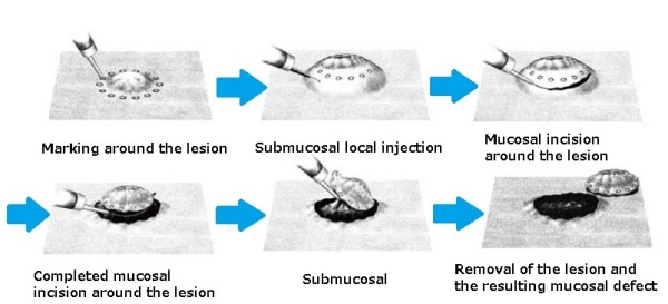
Fig. 1. Endoscopic submucosal dissection (ESD)

Photo 1. Examples of ESD
Around 2000, endoscopic submucosal dissection (ESD) procedure (Fig. 1 and Photo 1) was developed in Japan. As one of the advanced institutes, the University of Tokyo Hospital was deeply engaged in this technical development. We estimate that this procedure is currently performed in ~50,000 cases every year in Japan for the esophagus, the stomach, and the colon combined. In this hospital, we performed 353 ESDs in the last fiscal year, and we still have one of the most cases in Japan. In the traditional treatment, an early-stage cancer was removed with a part of the organ by opening the abdomen or chest by surgical procedures. ESD, however, enables us to resect an early-stage cancer with an endoscope, while preserving the normal surrounding tissue of the organ. Except for Japan, there are almost no other countries in the world where ESD procedure can be safely performed throughout the country. Therefore, several physicians visit this hospital from abroad to watch and learn ESD every month.
ESD was developed at the Department of Gastroenterology, but our department was also used. This use triggered communication between gastroenterologists (whose strong skill is ESD) and gastrointestinal surgeons (whose strong skill is laparoscopic surgery). As a result, both of their strong skills were recently combined to develop a new surgical technique that originated from the University of Tokyo Hospital. This procedure is called non-exposed endoscopic wall-inversion surgery (NEWS) (Fig. 2 and Photo 2), and it is attracting strong global attention as a therapy that is a step ahead of ESD.
A gastric submucosal tumor is difficult to resect by ESD procedure, and also the traditional procedure for this tumor requires removal of a large part of the stomach; however, the new procedure NEWS enables us to resect only a minimal area of the stomach. Similarly, early-stage gastric cancers with a low possibility of lymph node metastasis and locally recurrent gastric cancers, therefore, are thought to be suitable lesions for NEWS. This procedure is a new technique that was approved for health insurance coverage April, 2014 and it is expected to be widely used in the entire Japan in the future. As seen above, the University of Tokyo Hospital has been leading the world in endoscopic medical care since the days of the development and practical application of gastric cameras.
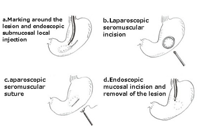
Fig. 2. Non-exposed Endoscopic Wall-inversion Surgery (NEWS)
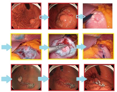
Photo 2. NEWS procedures
Treatment is performed with the cooperation of gastroenterologists and gastrointestinal surgeons (red-edged photos: gastroenterological procedures, yellow-edged photos: gastrointestinal surgical procedures).
a. Submucosal tumor of 20 mm in diameter on the upper anterior wall of the gastric body
b. Marking around the lesion on the mucosa
c. Marking around the lesion on the serosa
d. Submucosal local injection and laparoscopic seromuscular incision
e. Laparoscopic seromuscular suture while inverting the lesion into the pocket
f. After the seromuscular suture
g. Mucosal incision from inside the stomach to resect the entire layer
h. After the resection
i. Clip suture of the resected mucosal area and removal of the lesion through the mouth
![]()