Top > About Us > News Letter > World-leading Regenerative Medicine
World-leading Regenerative Medicine
—Focusing on surgical treatment for dental and oral-maxillofacial diseases
Message from the University of Tokyo Hospital to the World
Department of Oral-Maxillofacial Surgery, Dentistry and Orthodontics
At the Department of Oral-Maxillofacial Surgery, Dentistry, and Orthodontics, we provide various treatments focusing on surgical treatment for patients with diseases of the teeth, mouth, and jaws including congenital morphological defects such as cleft lip/cleft palate, jaw deformity, and injuries such as bone fracture, oral cancer, and infection. We also surgically reconstruct jawbone defects by inserting dentures or dental implants. Moreover, orthodontists correct malocclusions. We have directed special attention to advanced medical research and its clinical applications. In particular, in collaboration with the Department of Tissue Engineering, we have promoted the research and development of regenerative medicine of bone and cartilage and its clinical introduction (Figs. 1 and 2). Several representative examples have been included below.

Figs. 1 and 2
(1) Promotion of regenerative medicine in maxillofacial surgery and development of a new titanium mesh tray
Various surgical techniques for maxillofacial bone defects due to tumor resection or injury have been reported. Vascularized free fibula grafting has been increasingly used in particular for mandibular reconstruction requiring a long bone with sufficient blood flow. A surgeon needs to precisely adjust the shape of the bone graft, to ensure that it closely fits the grafting site during surgery. Because the maxillofacial region is complicated in shape and contour, a surgeon may find it difficult to achieve perfect restoration, which may ultimately affect patient satisfaction.
For such situations, we have recently introduced the bone restoration procedure using cancellous bone fragments collected from the ilium and a titanium mesh tray. In this procedure, the bone defect is filled with cancellous bone fragments and then covered with an appropriately shaped titanium mesh tray. This treatment enables the recovery of the natural shape and bone regeneration. Moreover, dentures or implants can be concomitantly inserted to restore proper occlusion. This regenerative approach may be ascribed to the body’s natural healing power.
Although this approach required sufficient experience to adjust and process the shape of the titanium mesh tray to that of the bone defect., recent development of a new mesh tray solved this problem. Unlike the conventional grid-like mesh tray, the new special hexagonal-pattern mesh tray allows quick 3-D processing without causing strain (Fig. 3).
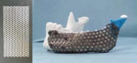
Fig. 3
(2) Development of a tailor-made bone (CT-Bone) and its clinical application
Autogenous bone grafting causes stress on the bone donor site, and the amount of bone that can be collected is limited. In other countries, the cadaver bones are frequently used for transplantation. However, artificial bones are commonly used in Japan, in order to avoid the risk of infection and ethical concerns.
In collaboration with the School of Engineering at the University of Tokyo, we have used a 3-D ink-jet printer to develop a method for producing an artificial bone (CT-Bone) that matched the shape of the original bone. We have used CT images to produce a 3-D plaster model and to design the shape of the bone graft using wax. We subsequently used a 3-D ink-jet printer to produce an artificial bone (CT-Bone) with a shape that matched the wax bone (Fig. 4).
Currently, the CT-Bone is used to treat patients with depression/malformation of the face. In these patients, the CT-Bone provides excellent compatibility with the original bones, and its rapid fusion with the original bones has been confirmed.
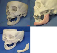
Fig.4
(3) Clinical application of the implanted regenerated cartilage
Generally, cartilage has a very limited capacity for self-repair. A large amount of cartilage cannot be harvested from other regions of the body. Therefore, there are great expectations for the introduction of regenerative medicine technology that enables the artificial production of cartilage in chondrocyte cultures. To date, liquid- or gel-based regenerated cartilage has been available. However, this type of regenerated cartilage is not sufficiently hard to be used for the repair of serious facial malformations. We subsequently developed regenerated cartilage that could be used to repair a nose with sufficient hardness and an optimal shape.
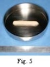
A small amount of ear chondrocyte is cultured with blood collected from the patient. The collagen and special material containing absorbable plastic (scaffolding material) are subsequently combined to produce the “regenerated cartilage—known as the “implanted” regenerated cartilage because it is “surgically implanted” rather than injected. We collect a small amount of cartilage from behind the patient’s ear. The small cut made during this treatment prevents any noticeable scars or deformations (Fig. 5).
We are the first team to use the implanted regenerated cartilage to treat the serious nasal deformation in a patient with cleft lip and palate. The regenerated cartilage can be created by culturing a small amount of cartilage, which is collected from a not easily visible site. This regenerative technique has been expected to contribute to the treatment of serious deformation.
Moreover, we believe that the technology of regenerated cartilage can be used in the treatment of the trachea and joints. To create the regenerated tracheal cartilage, the standard regenerated cartilage is processed into a curved plate. We plan to treat patients with long-term post-tracheotomy tracheal stenosis by inserting the wedge-shaped regenerated tracheal cartilage. We are in the process of developing a regenerated joint called the NeoJoint that has an optimal structure and strength to be implanted into the knee affected by osteoarthritis. Structurally, the NeoJoint is a combination of regenerated cartilage and artificial bone. Because we need many cells to produce the NeoJoint, we aim to determine whether induced pluripotent stem (iPS) cells can be used in the development of the NeoJoint.
In collaboration with the Department of Tissue Engineering, the Department of Oral-Maxillofacial Surgery, Dentistry, and Orthodontics progressively introduces world-leading regenerative medicine and advanced medical technology into the treatment of dental and oral-maxillofacial diseases.
You may have heard it, but…
Question: What is regenerative medicine?
Answer: Regenerative medicine is a medical therapy that will enable the body to regenerate a body part (tissue or organ), which no longer functions normally or has lost its function because of disease or injury. Japanese and overseas researchers are hard at work on the regeneration of various tissues and organs such as bones, skin, and vessels. Some research findings have already been applied clinically, including a method in which some cells or tissues collected from patients are cultured for autogenous transplantation. Recently, researchers have used stem cells—including iPS cells—to develop new regenerative medicine methods. Regenerative medicine has the possibility of treating diseases that cannot be treated using conventional methods. Moreover, regenerative medicine is expected to solve the problem of the shortage of organ transplant donors and organ rejection.
![]()